by Keith Benjamin | Nov 4, 2023 | Occular Anatomy
Layers of The Eye: Overview The human eye is a complex organ composed of several layers, each with a specific function. Here's a brief overview of these layers, starting from the outermost to the innermost: 1. Cornea and Sclera (Fibrous Tunic): Cornea: The transparent...
by admin | May 4, 2010 | Occular Anatomy
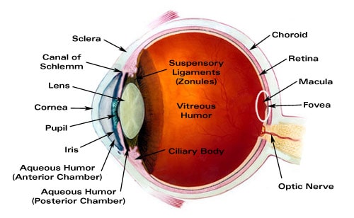
An outstanding overview of the major structures of the eye from the University of Missouri Medical School including: the Cornea, Lens, Ciliary Body, Iris, Ciliary Epithelium, Canal of Schlemm, Neuron Chain, Rods and Cones, Neural retina, and Fovea Centralis.
by admin | May 4, 2010 | Occular Anatomy
Extraocular Muscles The stabilization of eye movement is accomplished by six extraocular muscles attached to the eye via the sclera. The six muscles and their function are: Lateral rectus - moves the eye outward, away from the nose Medial rectus - moves the eye...
by admin | May 4, 2010 | Occular Anatomy
Refractive errors occur when abnormalities of the eye prevent the proper focus of light on the retina. Emmetropia refers to an eye free of refractive errors. Common Refractive Errors Two common types of refractive errors are myopia and hyperopia. Myopia Myopia, also...
by admin | May 4, 2010 | Occular Anatomy
Amblyopia - a reduction or dimming of vision in an eye that appears to be normal. Also commonly known as lazy eye, amblyopia is an eye condition noted by reduced vision not correctable by glasses or contact lenses and is not due to any eye disease. The brain does not...


