Layers of The Eye: Overview
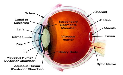
The human eye is a complex organ composed of several layers, each with a specific function. Here's a brief overview of these layers, starting from the outermost to the innermost:
1. Cornea and Sclera (Fibrous Tunic):
-
- Cornea: The transparent front part of the eye that covers the iris, pupil, and anterior chamber. It refracts light and is responsible for a significant portion of the eye's focusing power.
- Sclera: The white part of the eye, a tough, fibrous tissue that extends from the cornea and covers the entire eyeball, except the part covered by the cornea. It provides protection and form.
- 2. Choroid, Ciliary Body, and Iris (Vascular Tunic or Uvea):
-
- Choroid: A vascular layer containing connective tissue; it lies between the retina and the sclera. The choroid provides oxygen and nutrients to the outer layers of the retina.
- Ciliary Body: A structure containing the ciliary muscle and ciliary processes; it connects the iris to the choroid. The ciliary body controls the shape of the lens and secretes aqueous humor.
- Iris: The colored part of the eye, which contains a muscle that adjusts the size of the pupil to regulate the amount of light entering the eye.
3. Retina (Nervous Tunic):
- Retina: The innermost layer of the eye, containing light-sensitive cells (rods and cones). It receives light and converts it into neural signals, which are sent to the brain through the optic nerve. The retina plays a crucial role in vision, enabling the perception of light, color, and detail.
Other key structures, although not layers, include:
- Lens: A transparent, biconvex structure located behind the iris, which focuses light onto the retina.
- Vitreous Humor: A clear gel that fills the space between the lens and the retina, helping maintain the eye's shape and optical properties.
Each layer and structure of the eye is essential for its overall function, working together to enable the complex process of vision.
Major Ocular Structures: In Depth
As mentioned above, the eye is made up of three layers: the outer layer called the (1) fibrous tunic, which consists of the sclera and the cornea; the middle layer responsible for nourishment, called the (2) vascular tunic, which consists of the iris, the choroid, and the ciliary body; and the inner layer of photoreceptors and neurons called the (3) nervous tunic, which consists of the retina.

The eye also contains three fluid-filled chambers. The volume between the cornea and the iris is known as the anterior chamber, while the volume between the iris and the lens is know as the posterior chamber, both chambers contain a fluid called aqueous humor. Aqueous humor is watery fluid produced by the ciliary body. It maintains pressure (called intraocular pressure or IOC) and provides nutrients to the lens and cornea. Aqueous humor is continually drained from the eye through the Canal of Schlemm. The greatest volume, forming about four-fifths of the eye, is found between the retina and the lens called the vitreous chamber. The vitreous chamber is filled with a thicker gel-like substance called vitreous humor which maintains the shape of the eye.
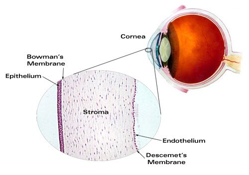
Light enters the eye through the transparent, dome shaped cornea. The cornea consists of five distinct layers. The outermost layer is called the epithelium which rests on Bowman's Membrane. The epithelium has the ability to quickly regenerate while Bowman's Membrane provides a tough, difficult to penetrate barrier. Together the epithelium and Bowman’s Membrane serve to protect the cornea from injury. The innermost layer of the cornea is called the endothelium which rests on Descemet's Membrane. The endothelium removes water from cornea, helping to keep the cornea clear. The middle layer of the cornea, between the two membranes is called the stroma and makes up 90% of the thickness of the cornea.

From the cornea, light passes through the pupil. The amount of light allowed through the pupil is controlled by the iris, the colored part of the eye. The iris has two muscles: the dilator muscle and the sphincter muscle. The dilator muscle opens the pupil allowing more light into the eye and the sphincter muscle closes the pupil, restricting light into the eye. The iris has the ability to change the pupil size from 2 millimeters to 8 millimeters.
Just behind the pupil is the crystalline lens. The purpose of the lens is to focus light on the retina. The process of focusing on objects based on their distance is called accommodation. The closer an object is to the eye, the more power is required of the crystalline lens to focus the image on the retina. The lens achieves accommodation with the help of the ciliary body which surrounds the lens. The ciliary body is attached to lens via fibrous strands called zonules. When the ciliary body contracts, the zonules relax allowing the lens to thicken, adding power, allowing the eye to focus up close. When ciliary body relaxes, the zonules contract, drawing the lens outward, making the lens thinner, and allowing the eye to focus at distance.
Light reaches its final destination at the retina. The retina consists of photoreceptor cells called rods and cones. Rods are highly sensitive to light and are more numerous than cones. There are approximately 120 million rods contained within the retina, mostly at the periphery. Not adept at color distinction, rods are suited to night vision and peripheral vision. Cones, on the other hand, have the primary function of detail and color detection. There are only about 6 million cones contained with in the retina, largely concentrated in the center of the retina called the fovea. There are three types of cones. Each type receives only a narrow band of light corresponding largely to a single color: red, green, or blue. The signals received by the cones are sent via the optic nerve to the brain where they are interpreted as color. People who are color blind are either missing or deficient in one of these types of cones.
Index of Refraction for Transparent Components of the Eye
Just as eyeglass lenses work to focus light where it is needed, so the different parts of the eye. Similarly, Just as different lens materials have different refractive indices, so do the different transparent parts of the eye.
Cornea: 1.37
Crystalline Lens: 1.42
Aqueous Humor: 1.33
Vitreous Humor: 1.33
Common Refractive Errors as They Relate to the Structures of the Eye
Refractive errors are vision problems that occur when the shape of the eye prevents light from focusing directly on the retina. The common refractive errors are myopia, hyperopia, astigmatism, and presbyopia, each relating differently to the structures in the eye:
- Myopia (Nearsightedness):
- Cause: Myopia occurs when the eyeball is too long relative to the focusing power of the cornea and lens, or when the cornea is too curved.
- Effect on Vision: This causes light rays to focus at a point in front of the retina, rather than directly on its surface, leading to blurred distant vision.
- Eye Structure Relation: The elongated shape of the eyeball or increased curvature of the cornea disrupts the normal focusing mechanism.
- Hyperopia (Farsightedness):
- Cause: Hyperopia is the result of the eyeball being too short, or the cornea having too little curvature.
- Effect on Vision: Light rays focus behind the retina instead of on it, making close objects appear blurry.
- Eye Structure Relation: The shorter-than-normal eyeball or less curved cornea shifts the focal point of light behind the retina.
- Astigmatism:
- Cause: This condition is caused by an irregular shape of the cornea or, occasionally, the lens inside the eye.
- Effect on Vision: It leads to blurred or distorted vision at all distances, as the irregularly shaped cornea or lens prevents light from focusing properly on the retina.
- Eye Structure Relation: The uneven curvature of the cornea or lens results in multiple focal points either in front of or behind the retina, or both.
- Presbyopia:
- Cause: Presbyopia is an age-related condition where the lens of the eye becomes less flexible, making it harder to focus on close objects.
- Effect on Vision: Difficulty in reading or seeing objects up close, usually noticeable after age 40.
- Eye Structure Relation: The reduced elasticity of the lens affects its ability to change shape (accommodate) for focusing on near objects.
How Eyeglass Lenses Help with Refractive Errors
Eyeglass lenses work by precisely altering the path and focus of light entering the eye to compensate for the specific refractive error, allowing the retina to receive a clear, focused image.
Optical wholesale labs work together with opticians and optometrists to produce lenses with the appropriate prescriptive correction that enables people with refractive errors to enjoy improved vision for both near and distant objects.
Visit OpticianWorks.com for comprehensive optician and NCLE study programs.
